Foot Muscles Mri : Ankle Mri Anatomy Youtube : Muscles of the foot are located on its rear and on the sole.
byAdmin-
0
Foot Muscles Mri : Ankle Mri Anatomy Youtube : Muscles of the foot are located on its rear and on the sole.. Musculoskeletal system | muscle structure and function. A magnetic resonance imaging (mri) was performed on a normal subject; More runners in the minimalist shoe group had increases in bone marrow edema than in the control. Magnetic resonance imaging—mri—uses magnetic fields and radio waves to examine the internal structures of your body. This article reviews the use of magnetic resonance imaging (mri) in the evaluation of the foot, including a discussion of bone and cartilage abnormalities in an article published in the august 2006 issue of this journal, the authors reviewed magnetic resonance imaging (mri) of the ankle.
It must be placed in the center of the magnet, to obtain homogeneous fat. Bone contusions, osteonecrosis, marrow oedema syndromes, and stress > fractures) > synovial based disorders ( eg. As a result, during walking the body's center of gravity normally fluctuates only 5cm in both vertical and lateral directions. Musculoskeletal system | muscle structure and function. More runners in the minimalist shoe group had increases in bone marrow edema than in the control.
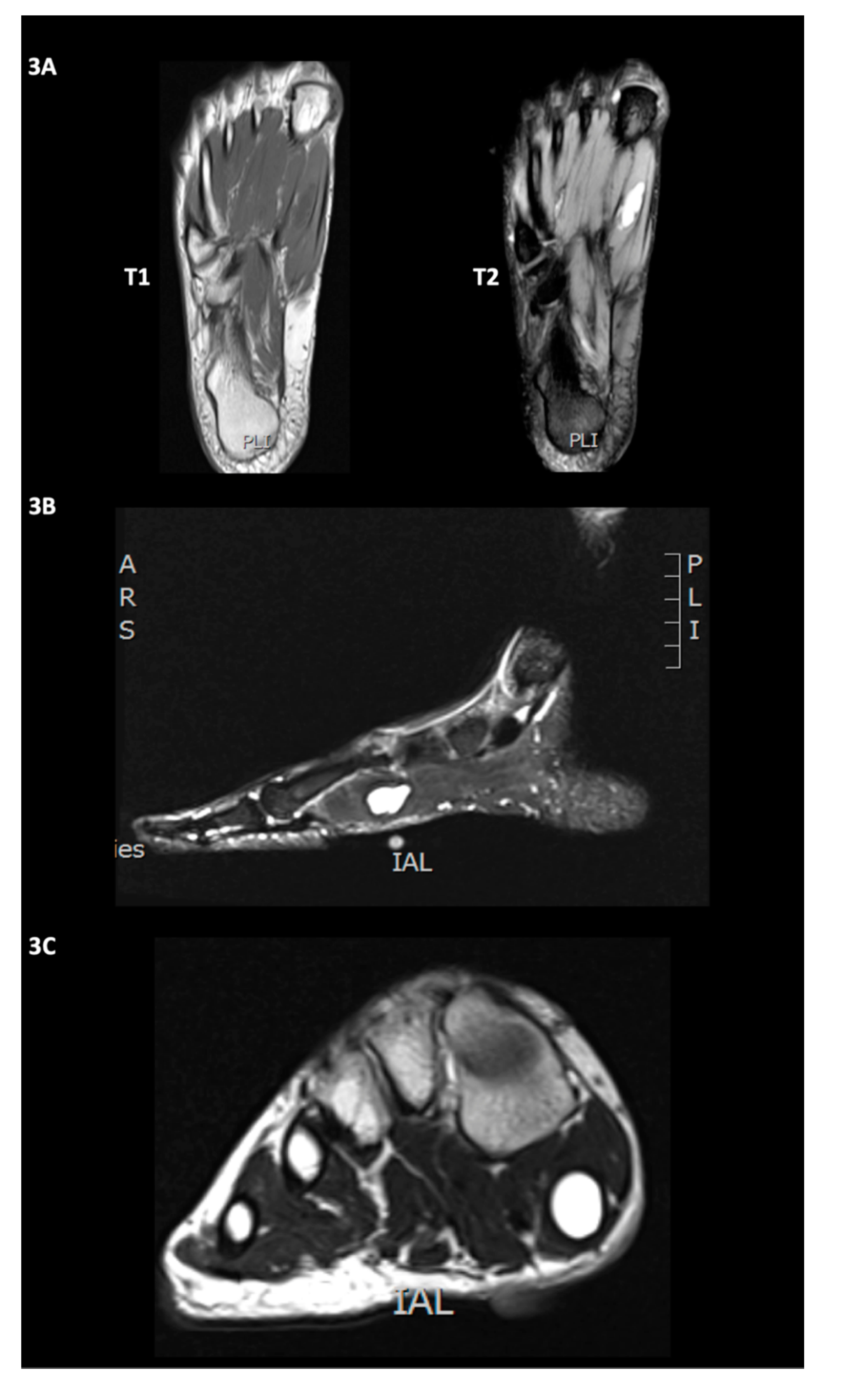
Diagnostics Free Full Text Intramuscular Ganglion Cyst Of The Flexor Hallucis Brevis Secondary To Muscle Tear A Case Report Html from www.mdpi.com Applications for magnetic resonance imaging (mri) of the foot and ankle disorders have expanded dramatically in the last decade.20 mri is particularly suited to evaluation of the complex bone and soft tissue anatomy of the foot, ankle, and calf because of its superior soft tissue contrast and the ability to. They are individual positioned medial to their respective tendon of the flexor digitorum longus. Posted by radiologyer at 8:12 am. Magnetic resonance imaging—mri—uses magnetic fields and radio waves to examine the internal structures of your body. This means that the little toe can only be extended by the extensor digitorum longus muscle only. There are 10 intrinsic muscles located in the sole of the foot. Interestingly the dorsal foot muscles generally have no insertion at the little toe. 12 photos of the foot muscle anatomy mri.
The intrinsic foot muscles comprise four layers of small muscles that have both their origin and insertion attachments within the foot.
This is a 30 year old with swelling on the lateral aspect of foot with evidence of soft tissue lesion in relation to the lateral aspect of the talus which appears isointense to the muscles on t1 and t2 weighted images & appears elongated extending from the anterosuperior calcaneum to the base of. Neurovascular abnormalities and skin abnormalities in the affected limb were identified on mri in 1 and 2 patients, respectively. The purpose of this study was to investigate the relationship of muscle mri findings and gait disturbance in myotonic dystrophy type 1 (dm1) patients. More runners in the minimalist shoe group had increases in bone marrow edema than in the control. The intrinsic foot muscles (ifm) are the main local stabilizers of the foot and are part of the active and neural subsystems that constitute the foot core. The flexor digiti minimi brevis (flexor brevis minimi digiti, flexor digiti quinti brevis) lies under the metatarsal bone on the little toe, and resembles one of the interossei. Epidemiology of tuberculosis etiology tuberculous spondylodiscitis clinical manifestations review of imaging findings: There can't be any metal in the room, not just where you have the mri. They act collectively to stabilise the arches of the foot, and individually to control movement of the digits. 12 photos of the foot muscle anatomy mri. ► shoulder ► elbow ► wrist ► finger ► thumb. Posted by radiologyer at 8:12 am. Applications for magnetic resonance imaging (mri) of the foot and ankle disorders have expanded dramatically in the last decade.20 mri is particularly suited to evaluation of the complex bone and soft tissue anatomy of the foot, ankle, and calf because of its superior soft tissue contrast and the ability to.
Perform routine foot plus coronal fmpspgr fat saturated pre and post gad images and axial post gad fmpspgr fat saturated images. The muscles acting on the foot can be divided into two distinct groups; 12 photos of the foot muscle anatomy mri. A magnetic resonance imaging (mri) was performed on a normal subject; Foot ulceration can subsequently lead to infections, such as cellulitis and osteomyelitis, and this may eventually the mri examination includes special attention for positioning of the foot.
Congenital Unilateral Hypertrophy Of The Foot Intrinsics A Rare Case And Review Of Literature Journal Of Orthopaedic Case Reports from www.jocr.co.in This is a 30 year old with swelling on the lateral aspect of foot with evidence of soft tissue lesion in relation to the lateral aspect of the talus which appears isointense to the muscles on t1 and t2 weighted images & appears elongated extending from the anterosuperior calcaneum to the base of. There are 10 intrinsic muscles located in the sole of the foot. Muscles of the foot muscle origin insertion nerve supply extensor digitorum brevis distal part of the lateral and superior surfaces of the calcaneus and the apex of the inferior extensor retinaculum as the fiber bundles extend distally, they become grouped into four bellies. Involved early gray = muscle: The intrinsic foot muscles comprise four layers of small muscles that have both their origin and insertion attachments within the foot. Interestingly the dorsal foot muscles generally have no insertion at the little toe. Synovitis, tenosynovitis, bursitis, and ganglion cysts) > congenital and developmental conditions( eg.dysplasia, tarsal coalition). The flexor digiti minimi brevis (flexor brevis minimi digiti, flexor digiti quinti brevis) lies under the metatarsal bone on the little toe, and resembles one of the interossei.
► shoulder ► elbow ► wrist ► finger ► thumb.
Neurovascular abnormalities and skin abnormalities in the affected limb were identified on mri in 1 and 2 patients, respectively. Perform routine foot plus coronal fmpspgr fat saturated pre and post gad images and axial post gad fmpspgr fat saturated images. There can't be any metal in the room, not just where you have the mri. There are 10 intrinsic muscles located in the sole of the foot. 12 photos of the foot muscle anatomy mri. These muscles lengthen eccentrically during the stance phase of running before shortening at the propulsion phase. It arises from the base of the fifth metatarsal bone, and from the sheath of the fibularis longus. It begins with short tendon bundles on the medial surface of the calcaneus calcaneus, fleshy bundles on the lower retentive flexor. A magnetic resonance imaging (mri) was performed on a normal subject; Lateral and medial processes of calcaneal tuberosity, and band of connective tissue connecti. In addition, an image of all the muscles of the back and plantar part of the foot, all tendons and tendon ligaments, blood vessels and nerves are obtained. The intrinsic foot muscles (ifm) are the main local stabilizers of the foot and are part of the active and neural subsystems that constitute the foot core. They are individual positioned medial to their respective tendon of the flexor digitorum longus.
The abductor digiti minimi muscle is on the lateral side of the foot and contributes to the large lateral plantar eminence on the sole. The muscles acting on the foot can be divided into two distinct groups; The intrinsic foot muscles (ifm) are the main local stabilizers of the foot and are part of the active and neural subsystems that constitute the foot core. Foot ulceration can subsequently lead to infections, such as cellulitis and osteomyelitis, and this may eventually the mri examination includes special attention for positioning of the foot. They act collectively to stabilise the arches of the foot, and individually to control movement of the digits.
Mr Sean S Right Foot Mri Youtube from i.ytimg.com Perform routine foot plus coronal fmpspgr fat saturated pre and post gad images and axial post gad fmpspgr fat saturated images. Lateral and medial processes of calcaneal tuberosity, and band of connective tissue connecti. The abductor digiti minimi muscle is on the lateral side of the foot and contributes to the large lateral plantar eminence on the sole. 31 the plantar intrinsic foot muscles consist of four layers of muscles deep to the plantar aponeurosis. Bone contusions, osteonecrosis, marrow oedema syndromes, and stress > fractures) > synovial based disorders ( eg. The muscles of the dorsum of the foot are a group of two muscles, which together represent the dorsal foot musculature. This article reviews the use of magnetic resonance imaging (mri) in the evaluation of the foot, including a discussion of bone and cartilage abnormalities in an article published in the august 2006 issue of this journal, the authors reviewed magnetic resonance imaging (mri) of the ankle. Muscles of the foot are located on its rear and on the sole.
► shoulder ► elbow ► wrist ► finger ► thumb.
Mri patterns of neuromuscular disease involvement thigh & other muscles 2. Indications for foot mri scan. The muscles acting on the foot can be divided into two distinct groups; Musculoskeletal system | muscle structure and function. More runners in the minimalist shoe group had increases in bone marrow edema than in the control. The intrinsic foot muscles (ifm) are the main local stabilizers of the foot and are part of the active and neural subsystems that constitute the foot core. The muscles of the dorsum of the foot are a group of two muscles, which together represent the dorsal foot musculature. As a result, during walking the body's center of gravity normally fluctuates only 5cm in both vertical and lateral directions. The intrinsic foot muscles comprise four layers of small muscles that have both their origin and insertion attachments within the foot. ► shoulder ► elbow ► wrist ► finger ► thumb. Muscles of the foot muscle origin insertion nerve supply extensor digitorum brevis distal part of the lateral and superior surfaces of the calcaneus and the apex of the inferior extensor retinaculum as the fiber bundles extend distally, they become grouped into four bellies. There can't be any metal in the room, not just where you have the mri. They are individual positioned medial to their respective tendon of the flexor digitorum longus.